Managing Vertigo
The In-Ear Crystals Behind "The Spins"
"It does not matter what the body is doing. It only matters what the head is doing and where it's positioned."
Tonya Fuller, MSPT, has treated benign paroxysmal positional vertigo (BPPV) every working day for the better part of two decades. "Sometimes it's more prevalent, sometimes less," Fuller shared in her late September presentation. "But at least one person walking through the door every single day has BPPV."
Even so, when it happened to her, she didn't recognize it right away. She stood up one morning and fell back into bed. Her husband asked what was wrong; she said she was dizzy and got up anyway. In the kitchen, she kept drifting into the couch on her left, unable to walk a straight line. She sat for two hours wondering why it wasn't going away until, as she put it, "hey, silly, you do this for a living, you should try to figure it out."
That moment captures something important about BPPV itself: the most common cause of vertigo in the world doesn't always look like spinning. Fuller's own case involved no vertigo at all, just visual distortion on one side and a profound loss of balance. Founder of Dynamic Mobility & Balance Center in Englewood, Colorado, and a physical therapist for 25 years, Fuller has spent the last 20 specializing in vestibular disorders, earning her Certified Advanced Vestibular Physical Therapist credential. Her clinical philosophy rests on a single, repeated principle: symptoms can be vague and different in every patient, but the underlying mechanism (displaced crystals in an inner ear canal the size of a thumbnail) follows rules precise enough to diagnose and treat with real confidence.
A System the Size of a Thumbnail
The vestibular system itself sits inside the bony labyrinth, a hard container in the skull holding the membranous labyrinth (the actual sensory organs) supplied by the eighth cranial nerve. Three semicircular canals (anterior, posterior, and horizontal) sense angular acceleration, sending the brain constant updates about which direction the head is turning. Two otolithic organs, the saccule and utricle, detect linear motion and head tilt. "It's really an amazing system," Fuller said, "and the whole thing is the size of your thumbnail."
Inside those otolithic organs sits a jelly-like structure called the otolithic membrane, embedded with calcium carbonate particles clinicians call otoconia or, more simply, crystals. Those crystals are dense enough to respond directly to gravity, bending the hair cells beneath them and sending directional signals down the nerve to the brain. BPPV happens when those crystals dislodge from where they belong and drift into one of the semicircular canals, where they have no business being.
Two Forms, One Underlying Problem
According to Fuller's September 22 presentation, BPPV comes in two forms, and distinguishing between them shapes everything about treatment.
In canalithiasis, the far more common form, the crystals float freely within the endolymph fluid of the canal. When the head moves into a provoking position, gravity pulls the crystals to the lowest point of the canal, dragging the fluid with them and pulling on a sail-like structure called the cupula. That pull triggers an abnormal firing signal, producing the sensation of spinning. Once the crystals stop moving, typically within 60 seconds, the vertigo stops too, though lightheadedness or nausea may linger.
Cupulolithiasis is rarer and behaves differently: the crystals actually adhere to the cupula itself rather than floating freely, producing longer-lasting symptoms that don't resolve as the fluid settles. The distinction matters clinically because it changes which treatment will actually work, as a maneuver designed to flush free-floating crystals out of a canal does little for crystals stuck directly to the sensing structure itself.
To top it all off, not all canals are affected equally. The posterior canal accounts for the large majority of cases; the horizontal canal shows up in roughly 20 to 30% of cases in Fuller's own practice (some newer, less-verified research puts the figure closer to 40%); and the anterior canal is the rarest, at around 2%, largely because its near-vertical orientation makes it mechanically difficult for crystals to migrate into. "When in doubt, treat the posterior canal," Fuller said. "If you believe it's BPPV, you can't really tell where it's located — treat the posterior canal, because there's a good chance you're going to treat the right canal."

104657 Clinical Management of Concussion: Assessment, Rehabilitation, and Return to Activity
Concussion management has evolved significantly over the past decade, with growing evidence supporting early, individualized, and active rehabilitation over prolonged rest. This two-hour, evidence-based course is designed to provide rehabilitation professionals with the knowledge and clinical tools needed to confidently assess, treat, and guide patients through every stage of concussion recovery. Participants will explore the pathophysiology of concussion, current clinical...
Listening Before Testing
Before any positional testing begins, the subjective interview with the patient in question does most of the diagnostic work.
"You really want to be listening to the patient, and not just putting down 'patient comes in with dizziness', and then you move on," Fuller said. The details patients offer, like "I woke up and the room was spinning", "I can't bend down to get something out of the dryer", or "I can't rinse my hair in the shower without feeling like I'm going to fall," all point directly at positional triggers that distinguish BPPV from other causes of dizziness. The Dizziness Handicap Inventory (DHI) is the tool Fuller relies on most, since it asks pointed questions about whether looking up, bending over, or rolling over in bed makes symptoms worse.
One clinical detail is easy to miss and, in Fuller's telling, essential: "It does not matter what the body is doing. It only matters what the head is doing and where it's positioned." A patient who can't get into a full long-sitting position, or who has limited neck mobility, can still be accurately tested and treated. By moving the body around a fixed head position rather than assuming the standard setup is the only option.
Reading the Eyes to Find the Canal
Positional testing depends on watching nystagmus (involuntary eye movement) closely enough to determine which canal is involved and on which side.
The Dix-Hallpike test remains the gold standard for the posterior canal, though Fuller was careful to correct a common misconception from her own training: "While we all learned that the Dix-Hallpike tested every canal, it's really not always effective in testing the other two canals." The roll test and bow-and-lean test are used for the horizontal canal instead, and deep head-hanging for the anterior canal.
Testing both sides isn't optional, even when a patient's symptoms strongly suggest one side. Because of how the semicircular canals pair up across sides, a phenomenon called coplanar pairing, a problem in the left posterior canal can produce a response that looks, at first glance, like it belongs to the right anterior canal. Test only one side, and a clinician can walk away with the wrong diagnosis entirely. The direction and duration of the nystagmus itself tells the rest of the story: upbeating torsional movement toward the affected side indicates posterior canal involvement; pure downbeating nystagmus (rare, and often flagged as atypical BPPV) can suggest anterior canal involvement or a central disorder; horizontal nystagmus points to the horizontal canal.
104659 Vestibular Hypofunction: Treatment Strategies for the Therapy Professional
In this live, interactive webinar we will discuss one of the most common disorders affecting the vestibular system: vestibular hypofunction. Discussions will include vestibular anatomy and physiology, common causes and symptoms of unilateral and bilateral vestibular hypofunction, medical and clinical testing, and treatment strategies to improve function, support participation in daily activities, and optimize functional outcomes.
Treating the Canals
Canalith repositioning is the modern name for what most clinicians still think of as the Epley maneuver, a distinction Fuller finds almost amusing. "The only reason it's not called the Epley maneuver anymore," she explained in her September presentation, "was because Dr. Epley said if you don't use vibration on the mastoid bone, it is not the Epley maneuver." Research has since shown the vibration isn't clinically necessary, but out of deference to its inventor, the vibration-free version now carries a different name. It remains the gold standard for posterior canal canalithiasis, and the numbers support the reputation: 88% effective after a single treatment, 96% effective within a week.
The horizontal canal has its own signature treatment, the barbecue roll (named, unsurprisingly, for the sequence of 90-degree turns that carries a patient nearly full-circle from their back to their stomach and back around). The Gufoni maneuver offers an alternative for horizontal canal cases, using a shorter, simpler sequence of positions. For cupulolithiasis, where crystals are adhered rather than floating, different maneuvers (including the Semont and Semont+) are needed to dislodge them.
One older approach, the Brandt-Daroff exercise, developed in the 1980s before more targeted maneuvers existed, still has a place for patients who struggle to tolerate faster repositioning techniques. It works through habituation rather than direct crystal repositioning, repeated positional movement that, over time, resolves symptoms in roughly 85% of cases after three weeks of consistent practice.
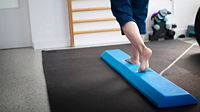
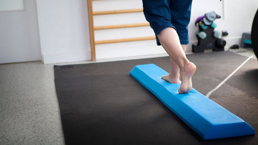
Post-treatment care matters as much as the maneuver itself. Patients are generally advised to sit upright for at least 15 minutes immediately afterward, and Fuller typically extends that recommendation to 30 to 60 minutes when patients are doing follow-up exercises at home. Residual dizziness, lightheadedness, or imbalance can persist even after successful repositioning, which is why Fuller treats balance function directly as part of recovery, often with foam-surface balance exercises, since an unstable surface forces greater reliance on the vestibular system itself rather than vision or proprioception to maintain balance.

104748 Manual Therapy and Corrective Exercises for Common Orthopedic Conditions
This course will develop manual therapy skills through advanced techniques combined with corrective functional exercises for common orthopedic upper and lower extremity conditions. Participants will explore multimodal approaches to facilitate personalized treatment plans that consider each patient’s activities and lifestyle. The course will focus on evidence-based manual therapy techniques and functional exercises designed to address difficult orthopedic conditions and support...
When the First Treatment Isn't the Last
BPPV has a reputation, in some clinical and insurance circles, as something that should resolve in one to three visits.
But, Fuller's own experience across 20 years of practice tells a more complicated story. "Some people's BPPV is very stubborn," she said. "Sometimes it shifts into a different canal. Sometimes it goes away and comes back the very next day." Recurrence is common, and cupulolithiasis can convert into canalithiasis over the course of treatment, which is why retesting at every follow-up, rather than assuming a prior diagnosis still holds, remains essential.
Fuller's guidance for clinicians facing a case that isn't resolving quickly is straightforward: if one maneuver isn't clearing the problem after repeated attempts, move to a different one rather than repeating the same approach indefinitely. With three distinct treatment options available for posterior canal involvement alone, and multiple options across the other canals and presentations, there's rarely a case that genuinely has nowhere left to go. Patients, she emphasized, should ultimately make a full recovery, it's simply a matter of matching the right maneuver to the right canal, and staying persistent until the crystals land back where they belong. ◼
Want to make the most of everything Impact CE has to offer?
The Ignite Membership unlocks Premium events and Masterclasses, Unlimited Live Webinars, Hundreds of Self-Paced Courses, a Growing Library of Recorded Content, and Free E-Library access — all in one place, for one low annual price. Learn more about Ignite today.
Resources
- Fuller, T. (2026, September 22). Managing vertigo: Strategies for BPPV [Webinar]. Impact CE.
- Bhattacharyya, N., Gubbels, S. P., Schwartz, S. R., Edlow, J. A., El-Kashlan, H., Fife, T., ... & Corrigan, M. D. (2017). Clinical practice guideline: benign paroxysmal positional vertigo (update). Otolaryngology–Head and Neck Surgery, 156(3_suppl), S1-S47.
- Bicknell, E. D., Ferguson, L., da Silva, A., Theoharidis, T., Gohil, K., Langford, J., ... & Bower, W. (2025). Routine benign paroxysmal positional vertigo (BPPV) physiotherapy management: a feasibility study. Age and Ageing, 54(3), afaf039.
- Edlow, J. A. (2025). Vestibular Rehabilitation in the Emergency Department—the Baby and the Bathwater. JAMA Network Open, 8(2), e2459518-e2459518.
- Hyo-Jung K, Park J, and Ji-Soo K. (2021). Update of benign paroxysmal positional vertigo. Journal of Neurology, 268, 1995-2000.
- Jeong S.H., Lee S.U., and Kim J.S. (2020). Prevention of recurrent benign paroxysmal positional vertigo with vitamin D supplementation: a meta-analysis. J Neurol, Aug 7, doi: 10.1007/s00415-020-09952-8.
- Lee, G. T., Agbelie, C. M. E., & Lee, T. H. (2025). Use of Physical Therapists in the Emergency Department for Benign Paroxysmal Positional Vertigo (BPPV): A Statewide Retrospective Analysis. Academic Emergency Medicine.
- Madrigal, J., Manzari, L., Figueroa, J. J., Castillo-Bustamante, M., & Figueroa, J. (2024). Understanding benign paroxysmal positional Vertigo (BPPV) and its impact on quality of life: a systematic review. Cureus, 16(6).
- Mohaddese, D.K., Ahadi, M., Maarefvand, M., and Jalaei, B. (2021). The Efficacy of the Half Somersault Maneuver in Comparison to the Epley Maneuver in Patients with Benign Paroxysmal Positional Vertigo. J Int Adv Otol, 17(5), 417 – 421.
- Wang, W. et al. (2020). New views on benign paroxysmal positional vertigo. Journal of Bio-X Research, 3, 78-82.